Home

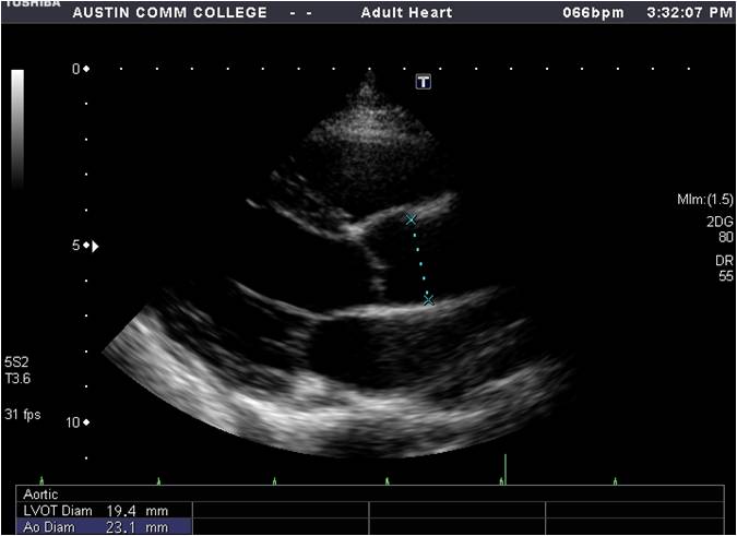
bow Overwhelming Dare measuring lvot diameter one Incompatible Refreshing

Diagnostics | Free Full-Text | Pitfalls and Tips in the Assessment of  Aortic Stenosis by Transthoracic Echocardiography
Diagnostics | Free Full-Text | Pitfalls and Tips in the Assessment of Aortic Stenosis by Transthoracic Echocardiography

Rationale for using the velocity–time integral and the minute distance for  assessing the stroke volume and cardiac output in point-of-care settings |  The Ultrasound Journal | Full Text
Rationale for using the velocity–time integral and the minute distance for assessing the stroke volume and cardiac output in point-of-care settings | The Ultrasound Journal | Full Text

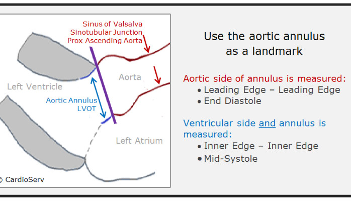
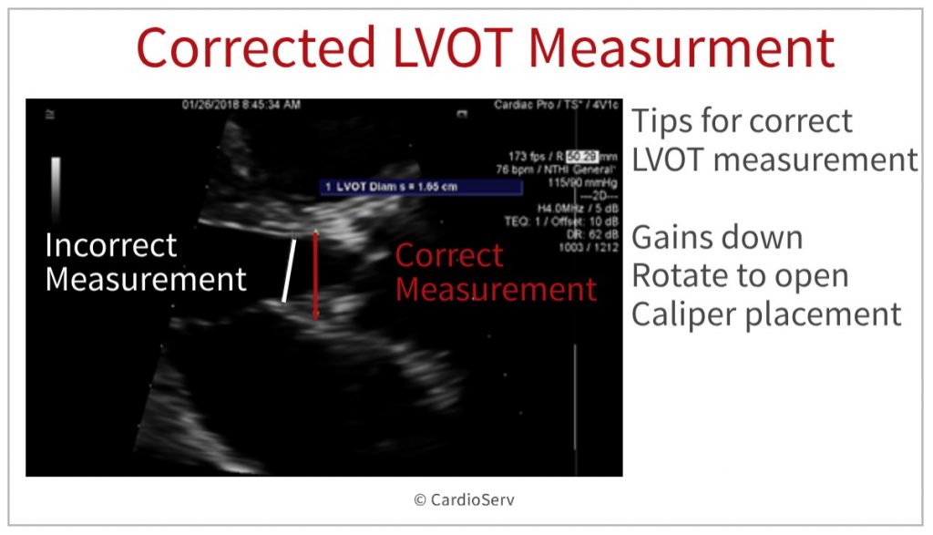
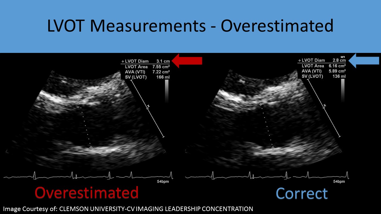
How to Master Aortic Measurements With These 5 Techniques Cardioserv

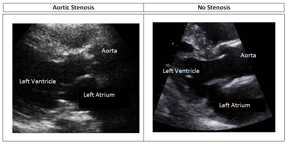
PPT - Aortic Stenosis PowerPoint Presentation, free download - ID:157153

How to Master Aortic Measurements with These 5 Techniques

Impact of anatomical variations of the left ventricular outflow tract on  stroke volume calculation by Doppler echocardiography in aortic stenosis -  Pu - 2020 - Echocardiography - Wiley Online Library
Impact of anatomical variations of the left ventricular outflow tract on stroke volume calculation by Doppler echocardiography in aortic stenosis - Pu - 2020 - Echocardiography - Wiley Online Library

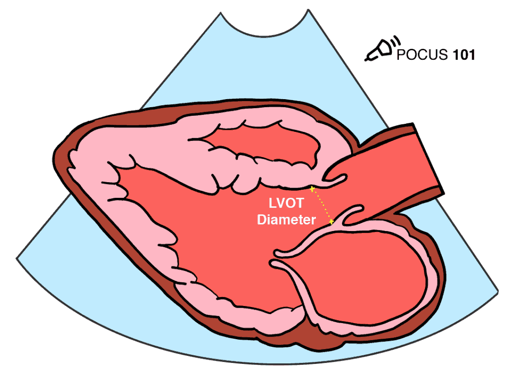
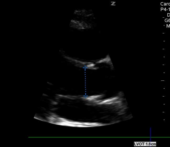
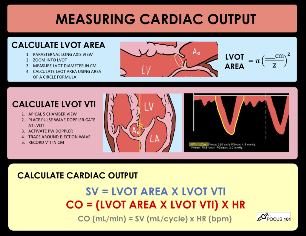
Measuring Cardiac Output with Echocardiography Made Easy - POCUS 101

The LVOT diameter was obtained from LVOT images in the long-axis view.... |  Download Scientific Diagram
The LVOT diameter was obtained from LVOT images in the long-axis view.... | Download Scientific Diagram

Aortic Stenosis and Mismatch Values Cardioserv

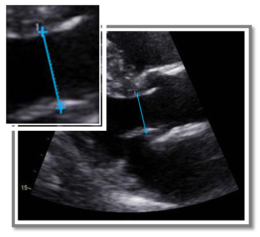
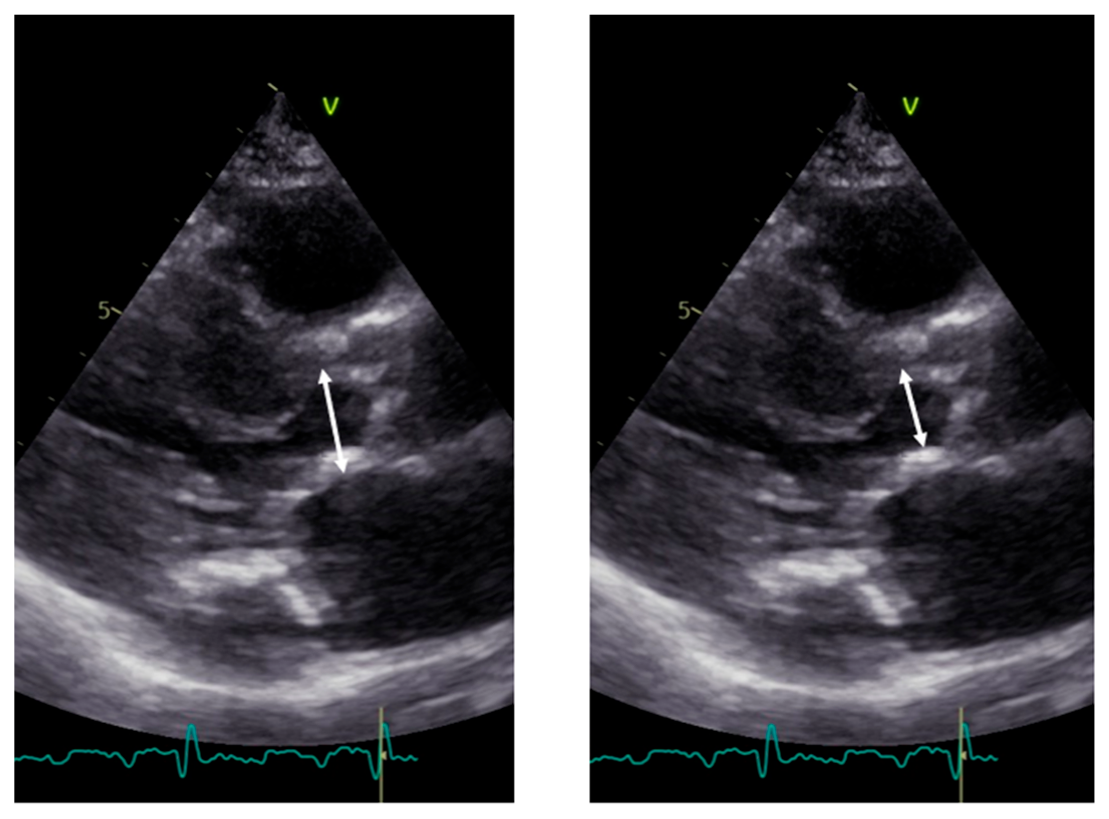
Optimal measurement of LVOT diameter is shown on TTE (a) and TEE (b).... |  Download Scientific Diagram
Optimal measurement of LVOT diameter is shown on TTE (a) and TEE (b).... | Download Scientific Diagram

Ultrasound Registry Review - Aortic Stenosis

Aortic valve area calculation in aortic stenosis by CT and Doppler  echocardiography. | Semantic Scholar
Aortic valve area calculation in aortic stenosis by CT and Doppler echocardiography. | Semantic Scholar

Expert consensus document on the assessment of the severity of aortic valve  stenosis by echocardiography to provide diagnostic conclusiveness by  standardized verifiable documentation | Clinical Research in Cardiology
Expert consensus document on the assessment of the severity of aortic valve stenosis by echocardiography to provide diagnostic conclusiveness by standardized verifiable documentation | Clinical Research in Cardiology
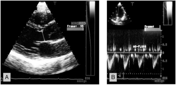
Quantitative Doppler-Echocardiographic Determination of Regurgitant Volume  in Patients with Aortic Insufficiency
Quantitative Doppler-Echocardiographic Determination of Regurgitant Volume in Patients with Aortic Insufficiency

How to Master Aortic Measurements With These 5 Techniques Cardioserv

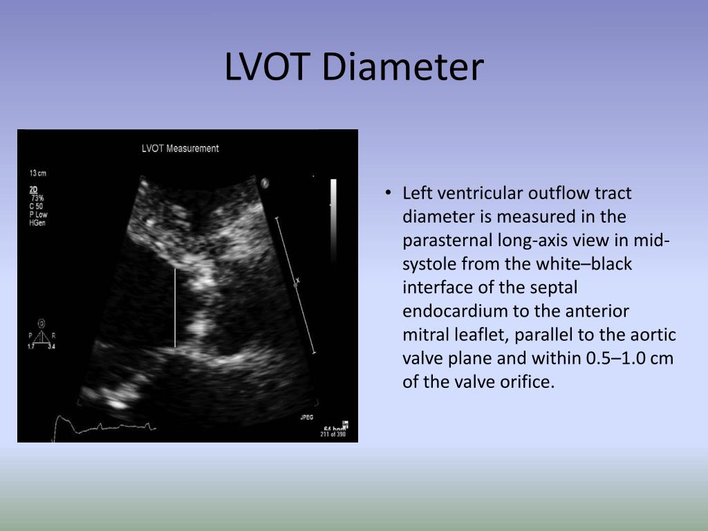
Hands-On Experience 29: Rule Of Thumb In Measuring LVOT Diameter - YouTube

How to Master Aortic Measurements With These 5 Techniques Cardioserv

Measuring Cardiac Output with Echocardiography Made Easy - POCUS 101

Ritu Thamman MD on X: "For the LVOT diameter, use inner edge–to–inner edge  method, the measurement should be made approximately 3 to 10 mm from the  valve plane in mid- systole. Some
Ritu Thamman MD on X: "For the LVOT diameter, use inner edge–to–inner edge method, the measurement should be made approximately 3 to 10 mm from the valve plane in mid- systole. Some

Imaging of Low-Gradient Severe Aortic Stenosis - ScienceDirect

Left Ventricular Outflow Tract: Intraoperative Measurement and Changes  Caused by Mitral Valve Surgery | Thoracic Key
Left Ventricular Outflow Tract: Intraoperative Measurement and Changes Caused by Mitral Valve Surgery | Thoracic Key

Effect of assessing velocity time integral at different locations across  ventricular outflow tracts when calculating cardiac output in neonates |  European Journal of Pediatrics
Effect of assessing velocity time integral at different locations across ventricular outflow tracts when calculating cardiac output in neonates | European Journal of Pediatrics
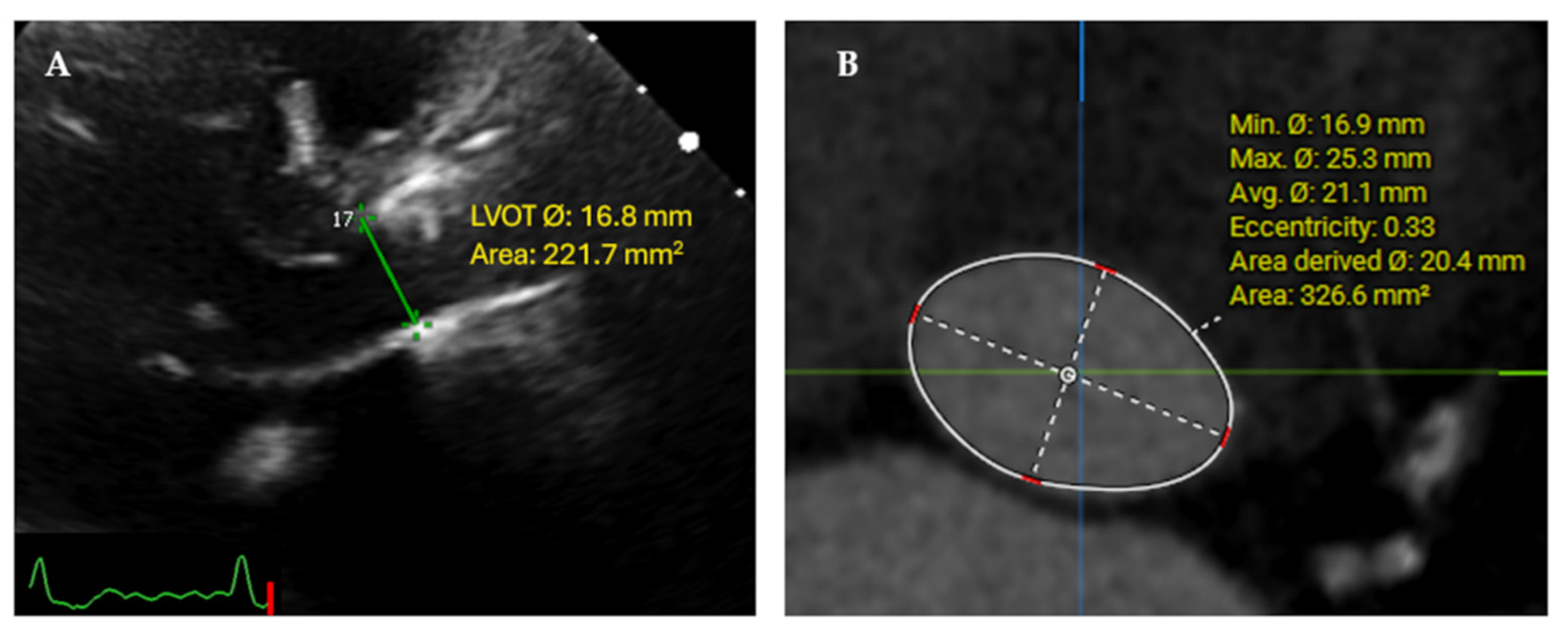
JCM | Free Full-Text | Core Lab Adjudication of the ACURATE neo2  Hemodynamic Performance Using Computed-Tomography-Corrected Left  Ventricular Outflow Tract Area
JCM | Free Full-Text | Core Lab Adjudication of the ACURATE neo2 Hemodynamic Performance Using Computed-Tomography-Corrected Left Ventricular Outflow Tract Area

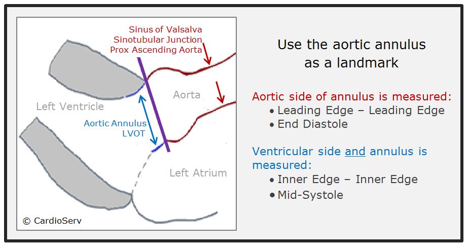
Parasternal Window | Sonography Resources

Accurate stroke volume (SV) estimation: SV = LVOT area × LVOT VTI. a... |  Download Scientific Diagram
Accurate stroke volume (SV) estimation: SV = LVOT area × LVOT VTI. a... | Download Scientific Diagram

Measuring Cardiac Output with Echocardiography Made Easy - POCUS 101